Point-of-care ultrasound (POCUS) is a critical extension of the physical examination in hospital medicine, and its use reflects a shift toward immediate, bedside-driven diagnostic decision making.1,2
While cardiac and pulmonary POCUS are now widely used, musculoskeletal (MSK) and vascular ultrasound remain underutilized despite their substantial clinical utility.3,4 MSK POCUS enables precise evaluation of soft tissue infections, joint effusions, tendon injuries, and procedural targets.3 Vascular POCUS informs fluid responsiveness, guides intravenous access, and assists in identifying deep vein thrombosis (DVT) with high sensitivity when performed by trained clinicians.4
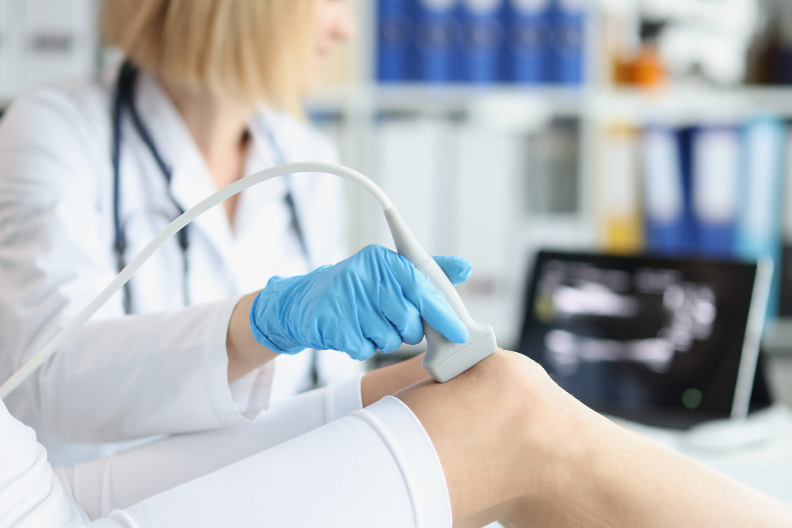
Case 1: The Diabetic Thigh Swelling
A 62-year-old man with poorly controlled type 2 diabetes presents with two days of progressive left thigh swelling, warmth, and erythema. The examination reveals diffuse tenderness but no clear fluctuance. The patient is febrile and tachycardic. Bedside MSK POCUS demonstrates a 2.5-cm hypoechoic fluid collection with posterior acoustic enhancement, confirming a drainable abscess layered beneath cellulitic tissue. Incision and drainage are performed at the bedside, followed by rapid clinical improvement.
Clinical Pearl
The physical exam alone is unreliable in differentiating cellulitis from abscess, especially in obesity, diabetes, or deep soft-tissue involvement.5 MSK POCUS improves diagnostic accuracy and allows earlier intervention for source control.5
Case 2: The Ambiguous Knee Effusion in Possible Septic Arthritis
A 44-year-old woman with long-standing rheumatoid arthritis presents with acute severe right knee pain and inability to bear weight. Despite swelling, the exam is obscured by habitus. MSK POCUS reveals a moderate suprapatellar effusion with synovial thickening. Ultrasound-guided arthrocentesis yields purulent joint fluid, leading to immediate antibiotic therapy and urgent orthopedic consultation.
Clinical Pearl
MSK POCUS reliably identifies even small or difficult-to-palpate joint effusions.6 Ultrasound-guided arthrocentesis increases first-pass success.7 Early recognition of septic arthritis is crucial; delays correlate with joint destruction and long-term functional impairment.8
Case 3: Shoulder Pain After a Hospital Fall
An older inpatient experiences a witnessed fall and subsequently develops severe lateral shoulder pain with limited abduction. Radiographs show no fracture or dislocation. MSK POCUS demonstrates an anechoic subacromial–subdeltoid bursal effusion and a partial-thickness tear of the supraspinatus tendon. This allows for targeted analgesia, early physical therapy intervention, and appropriate outpatient orthopedic follow-up without the need for urgent MRI.
Clinical Pearl
MSK POCUS can quickly differentiate soft-tissue injuries, guide management, and prevent unnecessary imaging.3 Ultrasound is uniquely positioned to visualize dynamic tendon motion and detect effusions or bursitis with no radiation exposure.9 Early identification of partial-thickness rotator cuff injuries can prevent chronic dysfunction and overuse of opioids.10
Case 4: Volume Status Dilemma in Acute Kidney Injury
A 76-year-old woman is admitted for pneumonia with acute kidney injury on labs. Exam findings are inconclusive, and her chart lists both prior heart failure and chronic diuretic use. Vascular POCUS shows a plethoric inferior vena cava with less than 15% collapsibility, and hepatic venous Doppler demonstrating systolic flow reversal—patterns consistent with venous congestion. Her diuretics are resumed rather than administering IV fluids, leading to improvement in renal function.
Clinical Pearl
Inferior vena cava assessment alone is imperfect, but integrating hepatic or portal venous Doppler markedly improves accuracy in evaluating venous congestion.11 POCUS-guided fluid management reduces iatrogenic volume overload, which is especially relevant in heart failure and renal failure.12 Recognizing venous congestion early prevents worsening acute kidney injury and respiratory compromise.13
Case 5: Suspected Femoral DVT With Delayed Imaging Availability
A 55-year-old man recovering from abdominal surgery develops right leg swelling and pain. Formal vascular ultrasound is delayed for 12 hours. A hospitalist performs a two-point compression ultrasound at the common femoral and popliteal veins, demonstrating noncompressibility of the right femoral vein. Anticoagulation is initiated immediately, and confirmatory imaging the next morning agrees with the bedside findings.
Clinical Pearl
Compression ultrasound performed by trained hospitalists has high sensitivity for proximal DVT.14 Early POCUS enables time-sensitive initiation of anticoagulation when formal imaging is unavailable or delayed.15 Recognizing proximal DVT promptly reduces morbidity associated with pulmonary embolism and post-thrombotic syndrome.16
Case 6: Difficult IV Access in a Septic Patient
A 47-year-old woman with sepsis and profound peripheral edema requires rapid IV access. Multiple blind attempts fail. Bedside vascular ultrasound identifies a 4-mm basilic vein at 0.6 cm depth. Real-time ultrasound guidance results in successful cannulation on the first attempt, allowing timely initiation of IV antibiotics and resuscitation.
Clinical Pearl
Ultrasound-guided peripheral IV access significantly improves success rates and reduces complications in patients with edema and obesity, and reduces the need for peripherally inserted central catheters and midline catheters in patients with vasculopathy.17 Consideration should be given to adopting ultrasound guidance as the default approach for difficult-access patients to increase chances of success while maintaining patient comfort.18

Dr. Asala
Dr. Abou Asala is a hospitalist and associate staff physician in the department of hospital medicine of the Cleveland Clinic Foundation, and a clinical assistant professor of medicine at the Cleveland Clinic Lerner College of Medicine, both in Cleveland.
References
1. Thind GS, et al. Point-of-care ultrasonography for the hospitalist. Cleve Clin J Med. 2021;88(6):345-359. doi: 10.3949/ccjm.88a.20141.
2. Bhagra A, et al. Point-of-Care ultrasonography for primary care physicians and general internists. Mayo Clin Proc. 2016;91(12):1811-1827. doi: 10.1016/j.mayocp.2016.08.023.
3. Situ-LaCasse E, et al. Utility of point-of-care musculoskeletal ultrasound in the evaluation of emergency department musculoskeletal pathology. World J Emerg Med. 2018;9(4):262-266. doi: 10.5847/wjem.j.1920-8642.2018.04.004.
4. Theophanous RG, et al. Point-of-care ultrasound screening for proximal lower extremity deep venous thrombosis. J Vis Exp. 2023;(192). doi: 10.3791/64601.
5. Subramaniam S, et al. Point-of-care Ultrasound for diagnosis of abscess in skin and soft tissue infections. Acad Emerg Med. 2016;23(11):1298-1306. doi: 10.1111/acem.13049.
6. Boniface K, et al. Point-of-care ultrasound for the detection of hip effusion and septic arthritis in adult patients with hip pain and negative initial imaging. J Emerg Med. 2020;58(4):627-631. doi: 10.1016/j.jemermed.2019.11.036.
7. Finnoff JT, et al. American Medical Society for Sports Medicine (AMSSM) position statement: interventional musculoskeletal ultrasound in sports medicine. Br J Sports Med. 2015;49(3):145-50. doi: 10.1136/bjsports-2014-094219.
8. Jeffers K, et al. What is the utility of point-of-care ultrasound for diagnosis of soft tissue abscess vs. cellulitis? J Emerg Med. 2025;72:121-128. doi: 10.1016/j.jemermed.2024.11.012.
9. Balaban M, et al. Evaluation of tendon disorders with ultrasonography and elastography. J Ultrasound Med. 2021;40(7):1267-1286. doi: 10.1002/jum.15520.
10. Dickinson RN, Kuhn JE. Nonoperative treatment of rotator cuff tears. Phys Med Rehabil Clin N Am. 2023;34(2):335-355. doi: 10.1016/j.pmr.2022.12.002.
11. Giangregorio F, et al. Assessing venous congestion in acute and chronic heart failure: a review of splanchnic, cardiac and pulmonary ultrasound: part 1: conventional B-Mode, colordoppler, and vexus protocol. J Clin Med. 2025;14(22):8147. doi: 10.3390/jcm14228147.
12. Patel S, et al. Objective methods of assessing fluid status to optimize volume management in kidney disease and hypertension: the importance of ultrasound. J Clin Med. 2023;12(19):6368. doi: 10.3390/jcm12196368.
13. Boyd JH, et al. Echocardiography as a guide for fluid management. Crit Care. 2016;20(1):274. doi: 10.1186/s13054-016-1407-1.
14. Fischer EA, et al. Hospitalist-operated compression ultrasonography: a point-of-care ultrasound study (HOCUS-POCUS). J Gen Intern Med. 2019;34(10):2062-2067. doi: 10.1007/s11606-019-05120-5.
15. Barrosse-Antle ME, et al. Point-of-care ultrasound for bedside diagnosis of lower extremity DVT. Chest. 2021;160(5):1853- 1863. doi: 10.1016/j.chest.2021.07.010.
16. Cox C, Roberts LN. Basics of diagnosis and treatment of venous thromboembolism. J Thromb Haemost. 2025;23(4):1185- 1202. doi: 10.1016/j.jtha.2025.01.009.
17. Galen B, et al. Reducing peripherally inserted central catheters and midline catheters by training nurses in ultrasound-guided peripheral intravenous catheter placement. BMJ Qual Saf. 2020;29(3):245-249. doi: 10.1136/bmjqs-2019-009923.
18. Vegas A, et al. Guidelines for performing ultrasound-guided vascular cannulation: recommendations of the American Society of Echocardiography. J Am Soc Echocardiogr. 2025;38(2):57-91. doi: 10.1016/j.echo.2024.12.004.