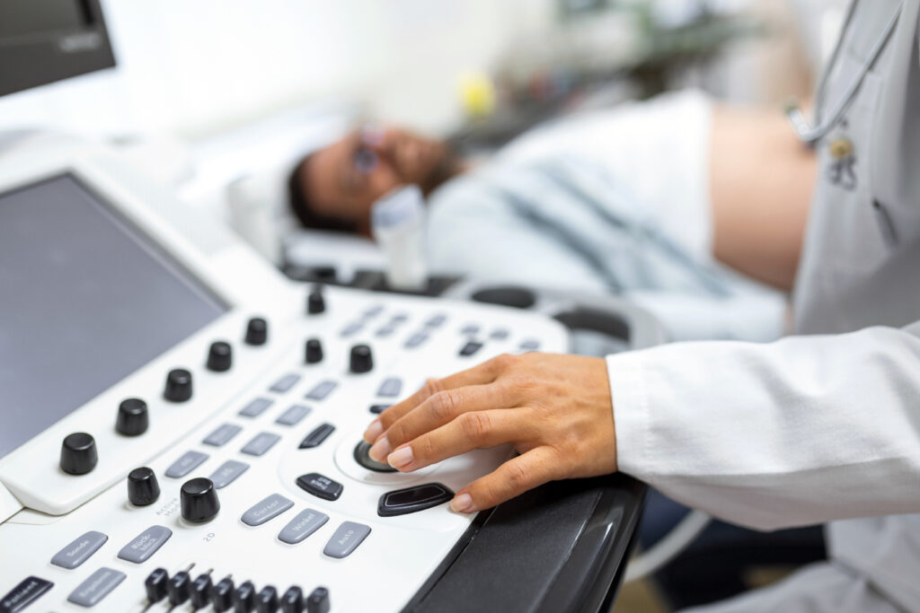
Abdominal ultrasound has become central in hospital medicine as a rapid, noninvasive, and radiation-free imaging modality for the evaluation of a wide range of acute and subacute abdominal conditions, in addition to its utility in evaluating volume status and offering procedural guidance.1,2
It plays an essential role in diagnosing pathologies in specific patient groups like children and pregnant women, where ultrasound is the preferred initial imaging test to address acute abdominal pain.3,4
The Society of Critical Care Medicine and the Infectious Diseases Society of America note that abdominal ultrasound is useful in critically ill patients with fever when abdominal symptoms or abnormal liver tests are present, aiding in the diagnosis of conditions such as acalculous cholecystitis, abscess, and appendicitis.5
Point-of-care ultrasound (POCUS) performed by hospitalists or non-radiologists has demonstrated moderate to excellent diagnostic accuracy for key abdominal pathologies after brief training and can alter management in a significant proportion of cases, with the diagnostic yield being highest when clinical suspicion is guided by history, physical examination, and laboratory findings.6-9
With that noted, operator skill and experience are critical determinants of diagnostic accuracy, and limitations include reduced sensitivity for certain pathologies and operator dependence.8,9
Case
A 65-year-old female with a history of end-stage renal disease secondary to focal segmental glomerulosclerosis presented to the emergency department for nausea that started earlier the same day. She had a renal transplant two years ago and is on immunosuppression.
On examination, Murphy’s sign was slightly positive. Labs showed stable kidney function with a high-normal white blood count. Computed tomography of the abdomen and pelvis showed questionable increased gallbladder wall thickness. A right upper quadrant ultrasound was ordered; however, it was not available overnight. The patient started reporting abdominal pain with worsening nausea and vomiting. Abdominal POCUS performed overnight by the nocturnist showed increased gallbladder wall thickness with pericholecystic fluid concerning for acute cholecystitis. Surgery evaluated the patient at the bedside, and the patient was taken to the operating room overnight for a cholecystectomy.
Clinical Pearl
Abdominal ultrasound serves as a convenient and effective tool to avoid delays in detecting life-threatening conditions, especially when resources are limited. This has been endorsed by multiple professional societies.10,11
Case
A 28-year-old patient with homozygous sickle cell disease was admitted for abdominal pain and found to be in a vaso-occlusive pain crisis requiring IV opioids and hydration. On hospital day two, the patient had an unwitnessed fall in the bathroom and subsequently developed acute, severe left upper quadrant abdominal pain with hypotension. The physical exam revealed abdominal guarding and rebound tenderness. POCUS performed by the hospitalist showed free fluid in Morison’s pouch and the splenorenal recess. An immediate CT of the abdomen confirmed hemoperitoneum with evidence of splenic injury. The patient was taken emergently to the operating room.
Clinical Pearl
POCUS is a rapid bedside tool that can reliably detect intraperitoneal free fluid when done with an experienced hand. In this case, it enabled prompt recognition of splenic rupture, expediting surgical management and ensuring patient safety.12,13
Case
A 45-year-old male with a history of pancreatic cancer with retroperitoneal metastasis was admitted to the hospital for intractable nausea and vomiting post-chemotherapy session. Abdominal pain was benign without tenderness or distention, so a CT was not pursued. Labs showed acute kidney injury presumed to be pre-renal due to low oral intake and volume loss with vomiting. The patient was started on aggressive IV hydration.
On his first night in the hospital, the patient started having worsening left flank pain. A CT was ordered, but was considered non-emergent, so radiology prioritized other CTs overnight with plans to take the patient for a CT in the morning.
POCUS was performed overnight at the bedside, revealing moderate left-sided hydronephrosis. Urology was consulted and boarded the patient in the early morning for obstructive uropathy with acute kidney injury.
POCUS can facilitate prompt and accurate diagnosis of urinary tract obstruction, enabling timely urology consultation and initiation of definitive management, enhancing patient safety and avoiding harm of delayed diagnosis.
Clinical Pearl
POCUS plays a central role in the rapid diagnosis of hydronephrosis at the bedside. It is highly sensitive to detect collecting system dilation and can be performed by non-radiologists with appropriate training, enabling timely identification of urinary tract obstruction. POCUS demonstrates sensitivity and specificity in the range of 85% to 90% for hydronephrosis, supporting its use as a first-line diagnostic tool to guide further management decisions.14,15
Case
A 62-year-old cachectic male with a history of heavy tobacco use presented with vague epigastric discomfort. A pulsatile abdominal mass was detected on exam. A CT was ordered but was considered non-emergent, and other CTs were prioritized overnight.
A bedside abdominal POCUS was performed by a hospitalist overnight, which revealed a 6.5 cm infrarenal abdominal aortic aneurysm. Due to these findings, a CT angiogram was done overnight and revealed an aneurysm with impending rupture. The finding prompted immediate vascular surgery overnight.
POCUS enabled rapid, non-invasive detection of a potentially life-threatening condition.
Clinical pearl
POCUS is highly effective in detecting abdominal aortic aneurysm, with sensitivity and specificity consistently reported in the range of 97% to 100% when performed by trained clinicians.16
In summary, abdominal POCUS is a rapid, noninvasive tool for the initial detection of cholecystitis, intra-abdominal free fluid, hydronephrosis, and abdominal aortic aneurysm.17 Limitations still exist, however, and include operator dependency and reduced sensitivity for subtle or early findings.18 While formal imaging remains necessary for equivocal or complex cases, POCUS serves as a handy tool in the initial evaluation, especially when resources are limited.19

Dr. Asala
Dr. Abou Asala is a hospitalist and associate staff physician in the department of hospital medicine of the Cleveland Clinic Foundation, and a clinical assistant professor of medicine at the Cleveland Clinic Lerner College of Medicine, both in Cleveland.
References
1. Thind GS, et al. Point-of-care ultrasonography for the hospitalist. Cleve Clin J Med. 2021;88(6):345-359. doi: 10.3949/ ccjm.88a.20141.
2. Fagley RE, et al. Critical care basic ultrasound learning goals for American anesthesiology critical care trainees: recommendations from an expert group. Anesth Analg. 2015;120(5):1041-1053. doi: 10.1213/ANE.0000000000000652.
3. Siegel MJ, et al. Ultrasonography of acute abdominal pain in children. JAMA. 1991;266(14):1987-9.
4. Zachariah SK, et al. Management of acute abdomen in pregnancy: current perspectives. Int J Womens Health. 2019;11:119-134. doi: 10.2147/IJWH. S151501.
5. O’Grady NP, et al. Society of Critical Care Medicine and the Infectious Diseases Society of America guidelines for evaluating new fever in adult patients in the ICU. Crit Care Med. 2023;51(11):1570-1586. doi: 10.1097/CCM.0000000000006022.
6. Wu X, et al. The accuracy of point-of-care ultrasound in the detection of gallbladder disease: a meta-analysis. Acad Radiol. 2024;31(4):1336-1343. doi: 10.1016/j. acra.2023.09.029.
7. González-Muñoz B, et al. Multi-organ clinical ultrasound as a complement to the diagnostic process in an internal medicine consultation. J Clin Ultrasound. 2024;52(7):837-845. doi: 10.1002/jcu.23710.
8. López Zúñiga MÁ, et al. Diagnostic capacity of pocket-sized ultrasound devices at point of care by a non-radiologist resident in patients with suspected abdominal pathology. Ultrasound Med Biol. 2020;46(2):263-268. doi: 10.1016/j. ultrasmedbio.2019.10.019.
9. Raman S, et al. Are we overusing ultrasound in non-traumatic acute abdominal pain? Postgrad Med J. 2004;80(941):177-9. doi: 10.1136/pgmj.2003.013805.
10. Boccatonda A, et al. Gastrointestinal ultrasound in emergency setting. J Clin Med. 2023;12(3):799. doi: 10.3390/ jcm12030799.
11. Laméris W, et al. Imaging strategies for detection of urgent conditions in patients with acute abdominal pain: diagnostic accuracy study. BMJ. 2009;338:b2431. doi: 10.1136/bmj.b2431.
12. Liu RB, et al. The practice and implications of finding fluid during point-of-care ultrasonography: a review. JAMA Intern Med. 2017;177(12):1818-1825. doi: 10.1001/ jamainternmed.2017.5048.
13. Leidi A, et al. Confidence and use of physical examination and point-of-care ultrasonography for detection of abdominal or pleural free fluid. A cross-sectional survey. Intern Emerg Med. 2022;17(1):113- 122. doi: 10.1007/s11739-021-02781-1.
14. Gaudreau-Simard M, et al. Imaging of medical patients with acute kidney injury: patterns of ultrasound use and the role of point-of-care ultrasound at a tertiary care center. J Gen Intern Med. 2025. Epub ahead of print. doi: 10.1007/s11606-025- 09704-2.
15. Nepal S, et al. Point-of-care ultrasound rapidly and reliably diagnoses renal tract obstruction in patients admitted with acute kidney injury. Clin Med (Lond). 2020;20(6):541-544. doi: 10.7861/ clinmed.2019-0417.
16. Mai T, et al. Point-of-care ultrasound performed by a medical student compared to physical examination by vascular surgeons in the detection of abdominal aortic aneurysms. Ann Vasc Surg. 2018;52:15-21. doi: 10.1016/j.avsg.2018.03.015.
17. Frankel HL, et al. Guidelines for the appropriate use of bedside general and cardiac ultrasonography in the evaluation of critically ill patients-part I: general ultrasonography. Crit Care Med. 2015;43(11):2479- 502. doi: 10.1097/CCM.0000000000001216.
18. López Zúñiga MÁ, et al. Diagnostic capacity of pocket-sized ultrasound devices at point of care by a non-radiologist resident in patients with suspected abdominal pathology. Ultrasound Med Biol. 2020;46(2):263-268. doi: 10.1016/j. ultrasmedbio.2019.10.019.
19. Arnold MJ, et al. Point-of-care ultrasonography. Am Fam Physician. 2020;101(5):275-285.